The Treatment of Pneumonia by Inhalation of Carbon Dioxide
Article in pdf-format >>
Published January 1930 in Archives of Internal Medicine
Yandell Henderson, PhD, Howard W. Harggard, MD, New Haven Connecticut and Pol N. Corrylos, MD, George L. Birnbaum, MD, New York with the collaboration of Ellen M. Radloff, BS, Cape Town, South Africa
I. The Relief of Atelectasis
The problem of pneumonia is peculiar. The pathogenic organisms involved are as well-known as those of typhoid or of diphtheria. Yet the mortality from such diseases as typhoid and diphtheria has been reduced far toward the vanishing point, while the mortality from pneumonia per hundred thousand of the population is almost the same as it was fifty years ago.
This lack of progress in the prevention and cure of pneumonia is the more noteworthy in view of the fact that the problem has been attacked as actively as that of other diseases by many able investigators using the methods of bacteriology, serology and preventive medicine. Thus the idea suggests itself that pneumonia may involve some factor that is not concerned in the other diseases and is not to be overcome by the methods effective against them. The factor peculiar to pneumonia may require treatment along a special line.
There is such a factor. It is one with which the internist is usually little concerned. For this reason it was long overlooked in pneumonia and has now only come to light through observations on the pneumonia following surgical operations. It is a factor with which surgeons are familiar; namely, occlusion of an infected organ and lack of drainage.
This new conception of pneumonia is that, as the infection takes place by way of the respiratory passages, it is of critical importance to keep. These passages open and drained. The bronchoscope has even been used for this purpose. According as the airways are closed or open, the infection is influenced to become acute or is ameliorated. This influence is exactly the same as that of the drainage or occlusion of a localized infection in any other part of the body.
Nature has provided the lungs with several protective devices and reactions. The most obvious is the cough reflex by which irritating foreign bodies are removed. Less obvious, but more constantly acting, are the movements of respiration which are probably accompanied by peristaltic contractions and relaxations of the air tubes. The mucosa lining these tubes bears cilia which produce a continual flow of secretion from the depths of the lungs outward.
Occlusion of an air tube puts all of these mechanisms for the clearing of the lungs out of action. The air in the occluded portion of the lung is soon absorbed, and the alveoli are deflated and collapsed. They are then gradually filled by accumulation of secretion. The conditions resulting are in all respects favorable to the development of microorganisms and, correspondingly, unfavorable both to the general defenses of the body and the special defenses of the lungs. It is a highly significant fact, as revealed by experiment, that in order to induce pneumonia in dogs it is not enough merely to introduce the pathogenic organisms into the lungs; it is essential also to narcotize the animals so deeply that the cough reflex is abolished and respiration is depressed. In general, depressed or shallow breathing tends to permit the development of pneumonia, and deep breathing with full ventilation of the lungs tends strongly to inhibit it.
Atelectasis as a factor in pneumonia
The critical importance of occlusion and collapse of the lung in the development of pneumonia might not have been discovered for a long time if investigation of another condition had not brought it to light. This was the occurrence of some degree of collapse of the lung in at least 10 per cent of all patients after surgical operations. The condition is seen particularly in patients who have undergone abdominal operations and whose breathing is thereafter impeded by the pain of the wound.
In tracing this matter still further it appears probable that the mechanism of collapse of the lung or, as it is better termed, atelectasis or apneumatosis (Coryllos), would not have been solved except for the fact that it was observed that this condition also results from the occlusion of a bronchus in diphtheria and from the plugging of a bronchus by a foreign body entering through the larynx and the trachea.
Thus, the evidence up to this point runs: Obstruction of a bronchus induces atelectasis. Atelectasis often follows surgical operations and is the condition precedent to postoperative pneumonia. Atelectasis gives to pneumonia the character of an occluded and undrained infection. This is the conception which investigation led Coryllos and Birnbaum (Ref 1) to formulate. Pneumonia is, in fact, as they define it, a "pneumococcic atelectasis."
Meanwhile, along other lines, the investigations of Henderson and Haggard resulted in the discovery (Ref 2) that inhalation of carbon dioxide is under several different clinical conditions, a preventive of pneumonia. The explanation of this effect is now found in the fact that deep breathing induced by inhalation of carbon dioxide dilates the lungs and thus prevents or relieves atelectasis. The principal conditions in which atelectasis and pneumonia are associated and in which inhalation of carbon dioxide is indicated as a preventive are as follows:
Atelectasis and pneumonia in the new-born
Before birth the lungs are normally atelectatic. Although the first cry is the sign that the lungs have been at least partially distended, it is fairly common for parts of the lungs to remain undistended for a considerable time. Such a continuance of atelectasis is recognized as predisposing to pneumonia. (Ref 3) To overcome a continuance or recurrence of the condition it is an ancient custom to make the child cry for fifteen minutes or more twice a day. The substitution of inhalation of 5 per cent carbon dioxide would be more in accord with the teachings of physiology. Such an inhalation has in fact been introduced by Henderson (Ref 4) as the most effective means of overcoming asphyxia in the new-born and has been widely adopted. It is probable that the routine use of this inhalation on all infants, those that breathe spontaneously, but often incompletely, as well as those which do not breathe without some additional stimulus, would go far to eliminate the occurrence of pneumonia during the first few weeks of life.
Post operative pneumonia
A death from pneumonia occurs now on the average, even in the best surgical practice, after each 240 major operations. (Ref 5) Nonfatal pulmonary complications occur frequently. It was shown, however, in papers of the highest importance for the development of this subject, by Scott and Cutler (Ref 6) in America and by Dzialoszinski with Meyer (Ref 7) and by Fisher (Ref 8) in Germany, that liability to postoperative atelectasis, massive, lobar or lobular, and to the pneumonia which frequently ensues, may be greatly reduced, indeed almost eliminated, by the routine administration of carbon dioxide at the termination of anesthesia and operation. That the benefit is due to the effects of carbon dioxide, rather than to the oxygen administered with it, is indicated by the fact that in German surgical practice the carbon dioxide is mixed merely with air without the addition of oxygen. The prophylactic effect of carbon dioxide in the prevention of postoperative pulmonary complications is clearly due to the full inflation of the lungs by the deep breathing induced by the inhalation.
It is probable that many previous observers have had premonitions on this matter; for Osier and McCrae (Ref 9) said: "The diagnosis of massive collapse of the lung after operation or injury may be difficult, especially [its differentiation] from lobar pneumonia. … In treatment, change in position . . . and encouraging the patient to breathe deeply help in prevention." Meltzer (Ref 10) had premonitions of this sort, but he was opposed to the conception, advocated in this country by Henderson, (Ref 11) of the physiologic and therapeutic control of respiration by carbon dioxide.
The inhalation of carbon dioxide in connection with anesthesia was introduced by Henderson, Haggard and Coburn (Ref 12) as a means of stimulating breathing, particularly for the purpose of inducing rapid elimination of the anesthetic after operation. An even more important result of such inhalation is the prevention of postoperative atelectasis and pneumonia.
Pneumonia after asphyxia
Experience has demonstrated that the treatment for carbon monoxide asphyxia by the inhalation of a mixture of oxygen and carbon dioxide, as introduced by Henderson and Haggard (Ref 13) a few years ago, not only is effective in the relief of asphyxia and the removal of carbon monoxide from the blood, but has resulted also in a notable decrease of postasphyxial pneumonia.
The occurrence of râles, edema, bronchopneumonia and lobar pneumonia in patients who have survived carbon monoxide asphyxia without the inhalational treatment has been noted by a number of observers. Their observations were summarized by Hamilton (Ref 14) in the statement that "evidence of injury to the lungs is shown in the fairly large proportion of the victims of gassing who have an excess of fluid in the respiratory tract." Drinker and Cannon (Ref 15) found in 1923, before the inhalational treatment was introduced, that the hospital records were incomplete regarding such cases, but that they indicate the probability that some degree of bronchopneumonia must be present in nearly all patients who do not succumb immediately following asphyxia by city gas. In contrast, among many hundreds of patients, of whose cases we have reports, treated in recent years with the inhalation of oxygen and carbon dioxide, not a single case of subsequent pneumonia has occurred.
In experiments on dogs asphyxiated with carbon monoxide it was found that animals resuscitated by the inhalation of carbon dioxide make a complete recovery without pulmonary complications. On the other hand, in animals that are not thus resuscitated the lungs are found at autopsy to be "edematous, and the alveoli to be filled with an almost homogeneous coagulum of albumin-rich fluid. Vascular stasis is also marked in some cases, although there is only a slight cellular inflammatory reaction and a few widely scattered small hemorrhages." (Ref 16)
No attempt will be made here to explain how asphyxia induces such a condition in the lungs. It is sufficient for present purposes to note the fact that, both in experiments on animals and in observations on patients, inhalation of carbon dioxide, simultaneously with the relief of asphyxia by means of oxygen, counteracts the tendency for this condition to give rise to postasphyxial pneumonia.
Pneumonia after asphyxia
Atelectasis, or apneumatosis (Coryllos), known in the first half of the last century and then almost forgotten, was described again in 1908 and 1910 by W. Pasteur (Ref 17) in cases of diphtheria with paralysis of the diaphragm. The clinical observations of Jackson and Lee (Ref 18) showed that bronchial obstruction by preventing the ventilation of a part of the lung permits a gradual absorption of the air within its alveoli and thus results in the deflation and collapse of a lobe or lobules or of the whole of one lung. More than fifty years ago, experimental evidence was obtained by Mendelsohn (Ref 19) and by Lichtheim (Ref 20) that the insertion of a plug in a bronchus induces atelectasis in the portion of the lung to which it leads. But only recently has this subject been developed on the basis of thorough experimental investigation by Coryllos and Birnbaum. (Ref 21)
They have devised plugs consisting of small rubber bags which are inserted in one of the main bronchi or in a branch by means of a bronchoscope and inflated so as to block off the airway completely. The dogs are narcotized with iso-amyl-ethyl barbituric acid to prevent coughing. Unilateral atelectasis develops in a few hours and is demonstrated by means of x-ray pictures. The most striking and most reliable index of the degree of atelectasis is afforded by displacement of the heart and mediastinum toward the side affected. The diaphragm is also elevated on that side and lowered on the side of the normal lung. In the dog the displacement of the heart is much greater than is possible in man, owing to the more rigid mediastinum in man, while the distortion of the diaphragm is greater in man than in the dog.
Following up this study of the effects of mechanical obstruction of the airways, Coryllos and Birnbaum (Ref 2) showed that lobar pneumonia is essentially a pneumococcic atelectasis. In their experiments, dogs which are deeply narcotized with iso-amyl-ethyl barbituric acid receive through the bronchoscope into one bronchus and its branches a small volume of a virulent culture of pneumococci concentrated by the centrifuge from a larger volume. The next day, a unilateral pneumococcic atelectasis with marked displacement of the heart toward the affected lung is found. This condition develops into a typical unilateral pneumonia with consolidation and hepatization. In the large majority of cases the disease terminates fatally in from one to three days.
Coryllos and Birnbaum also correlated this experimental evidence with clinical observations demonstrating that atelectasis is a critical factor in pneumonia. Thus, both experimental and clinical evidence indicates that in the development of pneumonia the first stage after infection is a catarrh which plugs the airways, great or small, with thick, sticky secretion and prevents their ventilation by the movements of respiration. The normal drainage is thus stopped, the air is absorbed, the lung or lobe or lobules are collapsed and an occluded area of infection is produced. In such areas, and only in such areas, pneumonia develops. The condition is sometimes successfully relieved in patients by aspirating the bronchi through a bronchoscope.
In pneumonia the heart is not displaced toward the normal lung, as it would be if the current conception were correct. In medical pneumonia, as in postoperative pneumonia, the heart is always displaced toward the pneumonic lung. This fact has now been observed also in pneumonia in children by Tallerman and Jupe. (Ref 22)
Experimental work
The Relief of Atelectasis.—The first series of experiments to be reported here was made for the purpose of determining the effectiveness of the inhalation of carbon dioxide in redilating a lung that had been collapsed by simple obstructive atelectasis without infection. In these experiments, twenty-four large dogs, from 12 to 25 Kg. in weight, were vised. They received, by intraperitoneal injection,
0.5 cc. per kilogram of a freshly made 10 per cent solution of iso-amyl-ethyl barbituric acid. Within fifteen minutes this induced, in nearly all cases, a deep narcosis lasting from twelve to twenty-four hours. An x-ray picture was taken to make sure that the heart and lungs were in normal condition and position. By means of a bronchoscope a rubber bag, similar to those used by Coryllos and Birnbaum but somewhat larger and fitted with a valve from a bicycle tire, was then inserted into the right bronchus and inflated with water so as to shut off as nearly as possible all the branches of the bronchus. In some of the earlier experiments a strong solution of sodium bromide or sodium iodide was used to inflate the bag; but it was found that if any of this solution escaped into the lung, it caused acute irritation or even immediate death. Water produces no such effect.
As soon as the bronchial plug was in place, the dog was laid in a special holder similar to that used by Coryllos and Birnbaum, so that its position could be adjusted as precisely as possible, and an x-ray picture was taken. The animal was then laid on its right side on the floor, where it remained in profound narcosis for many hours.
The next day, a second x-ray picture was taken and showed in all cases a degree of atelectasis and of displacement of the heart corresponding to the number of branches of the bronchus occluded; for atelectasis develops only in an area the airway of which is completely shut off. With the dog under ether anesthesia and by means of the bronchoscope the plug was withdrawn from the bronchus and the animal was placed in an atmosphere of air to which from 5 to 7 per cent of carbon dioxide was added. For this purpose a chamber 12 by 9 feet and 7 feet high (3.6 by 2.7 by 2.1 meters) was used. Its walls consisted of window sash; the joints were made airtight with soft asphalt. Beneath the wooden floor and above the wooden ceiling were layers of soft asphalt. It had double doors to form an air lock and was cooled by a coil of pipes (automobile radiator) connected to the main water supply of the laboratory and draining into a sink in the chamber. Carbon dioxide gas was run into this chamber from a cylinder of liquefied "carbonic acid" through a meter and was mixed with the air by an electric fan. The contents of the chamber were determined at intervals by gas analysis with an Orsat analyzer. Usually the initial concentration was about 7 per cent and dropped to 5 or 6 per cent in the course of an hour or two. From four to six animals were put into the chamber together and were allowed to move about freely. They breathed deeply but appeared comfortable and sometimes slept.
After the animals had been in the chamber from thirty to sixty minutes they were removed from it and another set of x-ray pictures was taken. These pictures were in striking contrast to those taken prior to the period in the chamber. They showed a restoration of a practically normal condition in the thorax.
The observations of Coryllos and Birnbaum (Ref 21) in earlier series of experiments have shown that simple obstructive atelectasis, such as these cases presented, usually clears up only in the course of several hours. It is therefore highly significant that after being in this chamber and inhaling carbon dioxide for only thirty to sixty minutes—shorter periods would probably have been sufficient—the atelectasis was in all cases relieved and the heart was restored to its normal position. These observations confirm experimentally the similar clinical observations of Scott and Cutler. (Ref 6) The changes in the thorax, and particularly the displacement toward the atelectatic right lung of the line of the left border of the heart, are shown for six experiments in figure 1. The significant details of the experiments are given in the legend.

Figure 1
Explanation of Figure 1
Fig. 1.—Relief of atelectasis by inhalation of carbon dioxide (drawings traced from x-ray films): 1, outline of the normal thorax of a dog, weighing 15 Kg., showing the ribs, heart and diaphragm. Normal x-ray pictures were made from all the animals, but as all these pictures are essentially similar, only one is here reproduced. 2 A, the thorax of dog AJJ, weighing 15 Kg., twenty-two hours after obstruction of the right common bronchus. Note the displacement of the heart, particularly of the left border (at the right in the drawing) away from the normal lung and toward the atelectatic lung. In this and succeeding pictures, cloudy outlines are indicated by ++4-+. 2 B, after removal of the bronchial plug, the animal was kept in a chamber containing air and an average of 6 per cent carbon dioxide for fifty minutes. This picture was then taken. Note that the left border of the heart (at the right in the picture) has returned to nearly the normal position and that the right border of the heart (on the left in the picture) has a distinct outline through most of its extent and is in the normal position. 3 A, the thorax of dog AL, weighing 26 Kg., twenty-one hours after obstruction of the right common bronchus. The right lung is almost completely collapsed and the heart is displaced, so that its left border is nearly in the midline. The right side of the diaphragm is obscured and the left side is distorted downward in a manner characteristic of contralateral atelectasis. 3 B, after removal of the plug from the bronchus, the animal was kept under light ether anesthesia for twenty or thirty minutes during which time it struggled vigorously. This picture was then taken and shows that the atelectasis had almost completely cleared up, allowing the heart and diaphragm to return to their normal positions. The x-ray pictures, from which 3 A and 3 are traced, are reproduced in figure 2. 4 A, the heart of dog AJ, weighing 15 Kg., twenty-two hours after the right common bronchus was obstructed. The picture shows extreme atelectasis of the right lung with displacement of the heart and distortion of the diaphragm. 4 5, after removal of the plug from the bronchus, the animal was placed for forty-five minutes in 6 per cent carbon dioxide; this picture was then taken. It shows that the atelectasis had cleared up and that the heart and diaphragm had returned to their normal positions. 5 A, the thorax of dog AM, weighing 14 Kg., twenty-one hours after obstruction of the right common bronchus. There is atelectasis in the greater part of the right lung with displacement of the heart and distortion of the diaphragm. 5 B, after removal of the plug from the bronchus the animal was placed in a chamber with 6 per cent carbon dioxide for forty minutes. The picture shows an almost complete return of the heart and diaphragm to the normal position. 6 A, the thorax of dog AN, weighing 15 Kg., nineteen hours after obstruction of the right common bronchus. The extent of atelectasis in the right lung is indicated by the displacement of the heart and the distortion of the diaphragm. 6 B, after removal of the plug from the bronchus, the animal was placed for forty minutes in 6 per cent carbon dioxide. At the end of this time, as the picture shows, the atelectasis had cleared up and the heart and diaphragm had returned to their normal positions. 7 A, the thorax of dog AO, weighing 13 Kg., nineteen hours after obstruction of the right common bronchus. There is an extreme degree of atelectasis. 7 B, after removal of the plug from the bronchus, the animal was placed for forty minutes in 6 per cent carbon dioxide. As the picture shows, the atelectasis at the end of this time had cleared up and the heart and diaphragm had returned to their normal position.
As the bronchi are innervated by the vagi, three experiments were tried to determine whether these nerves may play a part in producing atelectasis. After the animal was narcotized, the right vagus was cut and the peripheral end stimulated; but no effects were observable in x-ray pictures after section or during stimulation. The bronchus was then plugged and atelectasis was developed precisely as in a lung with normal innervation. Next day, the atelectasis was relieved by the inhalation of carbon dioxide, precisely as in animals in which the nerve was not cut. These observations, so far as they go, indicate that the innervation of the lung is not involved in the production or relief of atelectasis, in agreement with Fontaine and Herrmann. (Ref 23)
Clear evidence that the main factor in redistending an atelectatic lung is deep breathing rather than the effects of carbon dioxide on the circulation, is afforded particularly by one experiment in which vigorous breathing was induced, after removal of the bronchial plug, by keeping the animal under light ether anesthesia for nearly half an hour. It struggled vigorously, and drew many deep breaths. In an x-ray picture taken immediately thereafter, the atelectasis had cleared up, while in the picture previous to this treatment the atelectasis was extreme. These pictures, shown in figure 2, afford conclusive evidence that it is deep breathing and not an effect of carbon dioxide on nervous or vascular mechanisms which clears up atelectasis.
The Relief of Pneumonia.—In this series of experiments, twenty-eight dogs were given the usual narcotizing dose of iso-amyl-ethyl barbituric acid. Prof. Leo F. Rettger supplied a virulent culture of pneumococcus type II from the Yale Laboratory of Bacteriology. A volume of 1.5 cc. of the culture per kilogram of body weight of the dog to be infected was centrifugated and decanted down to 0.2 cc. per kilogram of the animal's weight; the sediment was mixed in this volume. After insertion of the bronchoscope into the right bronchus, this concentrated suspension of pneumococci was insufflated into the bronchus and its branches.
After a preliminary group of experiments on six dogs, for practice in technic and procedure and for untreated controls, a series of experiments was carried out on twenty-two dogs. In this series, two dogs were found dead, chiefly from iso-amyl-ethyl barbituric acid poisoning, on the morning following narcotization, and four failed to develop pneumonia. Of the remaining sixteen all developed pneumonia—in most cases so severely that, judging by the six control animals and by the experience of Coryllos and Birnbaum, few if any would have survived. However, after spending various periods from two to twenty-four hours in from 5 to 7 per cent carbon dioxide in the chamber, all except three of the sixteen made a complete recovery. Even in those which died, autopsy showed that in two cases the cause of death was the suppurative pneumonitis, probably due to Bacillus coli, as was observed by Coryllos and Birnbaum, and as occurred also in some of our dogs a day or two after withdrawal of the bronchial plug in the preceding series of experiments.
More significant than such statistics, however, are the x-ray pictures, illustrations of which are given in figures 3 and 4, and outline drawings in figure 5. These figures and their legends show clearly: (1) that in all these cases of pneumonia, atelectasis was a striking feature, and (2) that under inhalation of carbon dioxide a reinflation of the pneumonic lung occurred in all cases. It is noteworthy, however, that while in all respects, except one, these experiments on pneumonia and its relief appear closely similar to the preceding experiments on atelectasis without infection, in this one feature there is a significant difference between the two sets of experiments. Different times are required. While considerably less than one hour in an atmosphere of carbon dioxide was sufficient to overcome simple atelectasis due to a bronchial plug, two hours, and in some cases twenty-four hours, in this atmosphere was required to reinflate the pneumonic lung.
During the period of inhalation of carbon dioxide the dogs with pneumonia drank, or if too weak to drink by themselves, were assisted in drinking, large amounts of water. This probably aided in their recovery, by overcoming anhydremia.
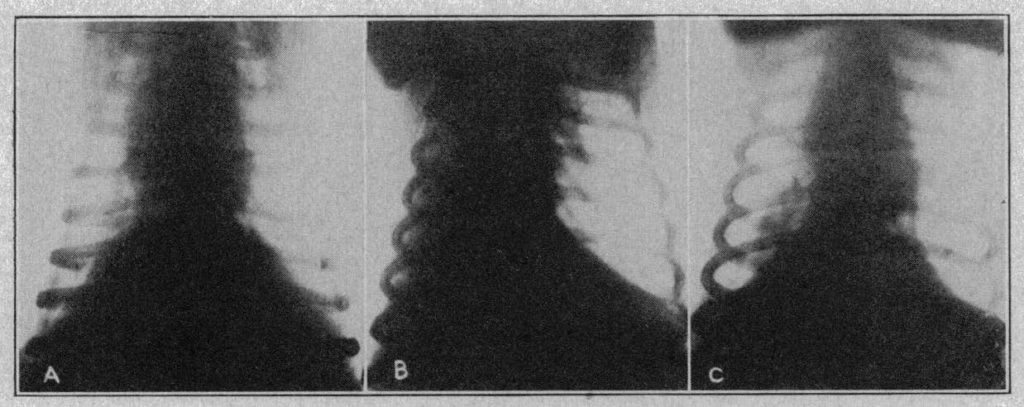
Fig. 2.—The thorax of dog AL: A, the normal condition, before the insertion of a plug in the right bronchus; B, extreme atelectasis the next day; C, half an hour later, reinflation of the right lung and restoration of the heart to the normal position, effected by removal of the plug and by a period of struggling and respiratory excitement under light ether anesthesia. The second and third of these pictures are also shown in outline as 3 A and in figure 1.
Comment
The observations reported in the two foregoing sections taken together throw light on two important matters: (1) the part which atelectasis plays in pneumonia, and (2) the effectiveness of the inhalation of carbon dioxide as a prophylactic and therapeutic measure against pneumonia.
In relation to the first of these topics the outstanding feature of the observations reported is the similarity of the experiments on pneumonia to those on simple obstructive atelectasis. Not only are the x-ray pictures of atelectasis from bronchial obstruction and pneumonia from intrabronchial infection strikingly alike, but—even more significant—the effects of the inhalation of carbon dioxide in the two series of experiments are closely similar. There can scarcely be any doubt as to the general character of the process involved in the opening up of a lung rendered atelectatic merely by bronchial obstruction without infection.
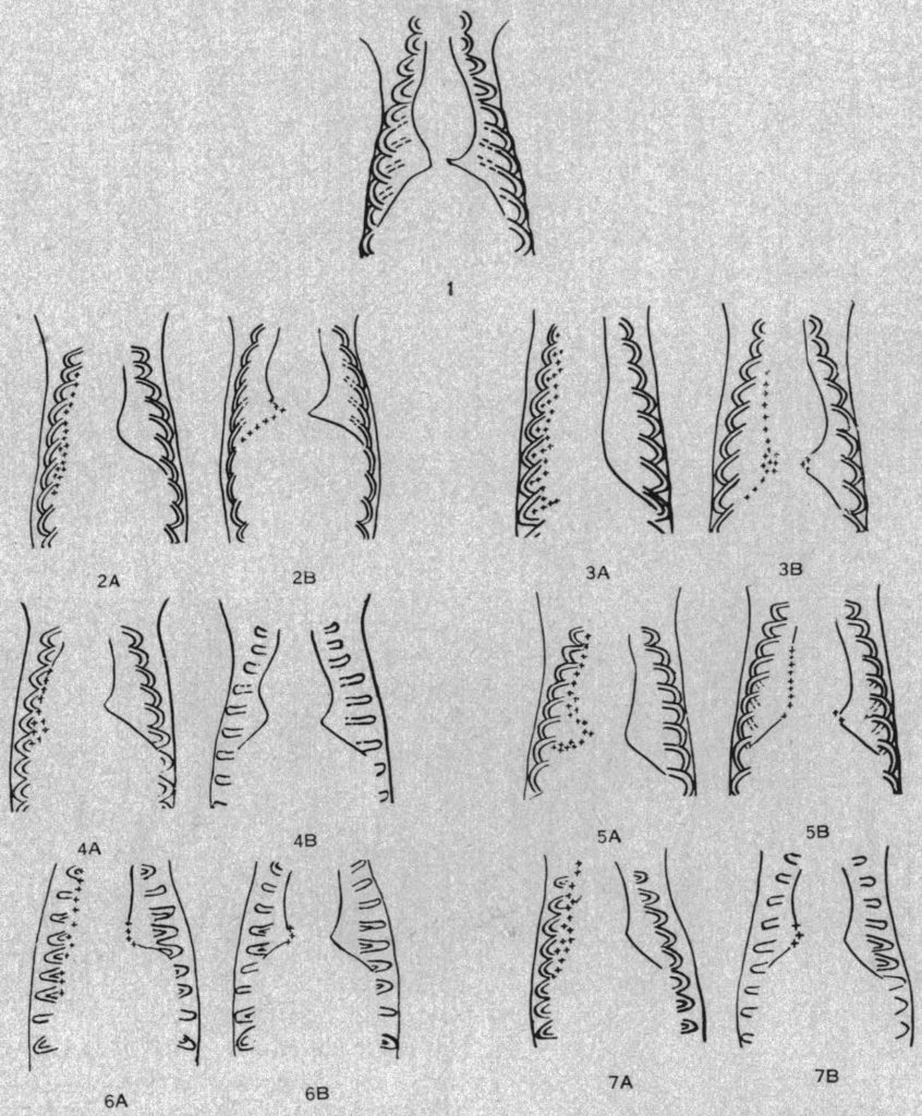
Fig. 3.—Relief of pneumonia by inhalation of carbon dioxide (drawings traced from x-ray films): 1, outline of the normal thorax of a dog, weighing 17 Kg., showing the ribs, heart and diaphragm. This will serve for comparison with the pictures of pneumonia indicated as A and of recovery indicated as in the subsequent pictures. 2 A, the thorax of dog PE, weighing 12 Kg., twenty-four hours after insufflation of 6 cc. of the sediment concentrated by the centrifuge from 18 cc. of a culture of pneumococci type II. Note the displacement of the heart toward the pneumonic lung, indicating extreme collapse of the right lung, which was nearly as opaque to x-rays as the heart was. In this and succeeding pictures, cloudy outlines are indicated by ++ ++· 2 B, the animal was placed in carbon dioxide, of an average concentration of 6 per cent, but showed only slight improvement during the first two hours. After twenty-three hours, however, the pneumonia had to a large extent cleared up and this picture was obtained. The animal made a complete recovery. It was killed four days later and when it underwent autopsy the lungs were found normal. The x-ray pictures, from which 2 A and 2 B are traced, are reproduced in figure 4. 3 A, the thorax of dog PF, weighing 17 Kg., twenty-four hours after insufflation of the sediment concentrated from 25 cc. of the culture of pneumococci. Note the extreme displacement of the heart; the left hand border is almost on the line of the vertebral column. A large part of the right lung was as opaque to x-rays as the heart itself. 3B, the animal was placed in carbon dioxide, average concentration of 6 percent, for two hours, after which this picture was taken. There is an almost complete restoration of the heart to the normal position, but an incomplete clearing up of the lung. The animal was extremely ill and died two hours later. At autopsy considerable areas in the right lung and a part also of the left lung were found to be hepatized. 4 A, the thorax of dog PG, weighing 20 Kg., twenty-four hours after insufflation of the sediment concentrated from 30 cc. of the culture of pneumococci. There is an intense unilateral pneumonia with displacement of the heart toward the infected lung. AB, the animal was placed in carbon dioxide, average concentration 6 per cent, but made only slight improvement during the first few hours. After twenty-four hours in decreasing concentrations of carbon dioxide from 7 to 4 per cent the picture was taken from which this drawing is traced. It shows that the right lung had largely cleared up, and that the heart had returned to nearly the normal position. The animal appeared entirely well the next day. It was killed and underwent autopsy four days later. The lungs were found normal except for a few small areas of congestion in the upper lobe of the left lung. 5 A, the thorax of dog PJ, weighing 17 Kg., twenty-four hours after insufflation of the sediment from 25 cc. of the culture of pneumococci. There is displacement of the heart to the right and opacity of the right lung except on its costal border. 5 B, the animal was placed in carbon dioxide, average concentration 6 per cent, for one and three-fourths hours. This picture was then taken and shows restoration of the heart to a nearly normal position, and a considerable degree of clearing in the right lung. The animal was found dead the next day. On autopsy it was found to have developed a suppurative pneumonitis with a strong fecal odor, probably due to infection by Bacillus coli. 6 A, the thorax of dog PS, weighing 14 Kg., twenty-three hours after insufflation of the sediment from 21 cc. of the culture of pneumococci. There is displacement of the heart to the right and complete opacity at the right lung except along the costal margin. 6 B, after two hours in carbon dioxide, average concentration 6 per cent, the heart had returned about half way to normal position and the right lung had cleared to a considerable extent, as shown. Before being placed in the chamber this dog showed a rectal temperature of 101 F. After two hours in carbon dioxide its temperature was 99.8 F. The animal appeared entirely well the next day, and was alive and healthy two weeks later. 7 A, thorax of dog PT, weighing 20 Kg., twenty-three hours after insufflation of the sediment from 30 cc. of the culture of pneumococci. There is displacement of the heart to the right, disappearance of the outline of the right side of the heart and of the right side of the diaphragm and a high degree of congestion in the right lung. 7 B, after two hours in carbon dioxide, average concentration 6 per cent, the heart had returned to nearly normal position and the right lung had largely cleared, as shown. The rectal temperature before the animal was placed in the carbon dioxide chamber was 102.8 F. At the end of the two hours of inhalation the temperature was 101.2 F. This dog made an uneventful recovery and was alive and well two weeks later.
Therefore the fact that inhalation of carbon dioxide results in a closely similar, even if much slower, opening up of a pneumonic lung affords conclusive proof that atelectasis is a factor in pneumonia. This conclusion is further reinforced by the fact that dogs with pneumonia, which would otherwise prove fatal, are generally restored to health as a consequence of this reopening of the lung. That pneumonia derives its virulent character to a large degree from the fact that it is an occluded infection is also thus proved; for the chief effect of opening up the pneumonic lung under inhalation of carbon dioxide is to provide drainage, although there may also be chemical and bactericidal effects, and influences on the circulation. Once it is reopened the lung may clear itself. As in many surgical infections, so in medical pneumonia, occlusion is the critical morbidic factor. Thus, the thesis of Coryllos and Birnbaum that lobar pneumonia is a pneumococcic atelectasis is completely confirmed.
On the second topic, the effectiveness of inhalation of carbon dioxide as a prophylactic measure against pneumonia, these observations are equally conclusive. The reason that the treatment of carbon monoxide asphyxia by inhalation of oxygen and 5 per cent carbon dioxide prevents a subsequent pneumonia—a fact which we have known for several years but could not previously explain—is now clear. Similarly, the discovery of Scott and Cutler and of German surgeons that inhalation of carbon dioxide at the termination of general anesthesia prevents post-operative pneumonia is confirmed and explained. The value of inhalation of carbon dioxide in the treatment of asphyxia and in connection with anesthesia is shown to be far greater than originally claimed by Henderson and Haggard. They introduced this treatment as a means of stimulating respiration and thus hastening the elimination of carbon monoxide, the anesthetics, and other foreign and toxic volatile substances from the blood. The accessory effect of preventing pneumonia after anesthesia and operation and after asphyxia is more important. If this inhalation proves even partially effective in the treatment of medical pneumonia, that result will become of yet greater importance.
Several hundred patients with pneumonia have been treated with oxygen and 5 per cent carbon dioxide by means of the inhalators employed in resuscitating patients with carbon monoxide asphyxia. In one group of 126 cases, the report shows that only nine patients died, while a number recovered by crisis immediately after the inhalation. Even more important than this low mortality is the fact that of the patients treated early nearly all recovered, while the deaths were nearly all among those treated late. In a much larger group of patients not so treated the mortality was 31 per cent. These observations are to be continued and will be reported in a later paper.
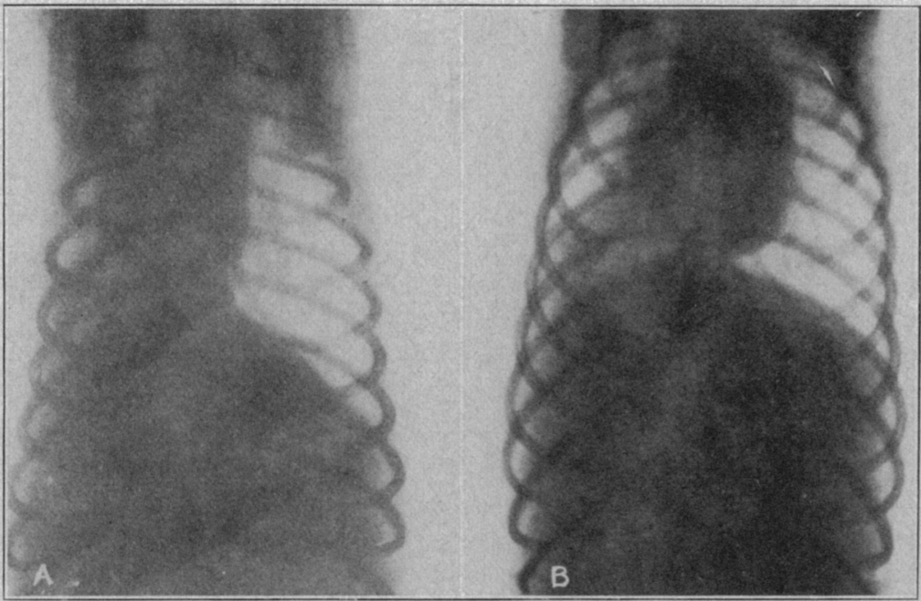
Fig. 4.—A shows unilateral pneumonia in dog PE, twenty-four hours after insufflation of pneumococci type II into the right bronchus ; B, restoration of the heart to the normal position and partial clearing up of the pneumonic right lung after twenty-three hours in 6 per cent carbon dioxide. This animal made a complete recovery. It was killed and underwent autopsy four days later; the lungs were found to be normal. These pictures are also shown in outline in 2 A and in figure 3.
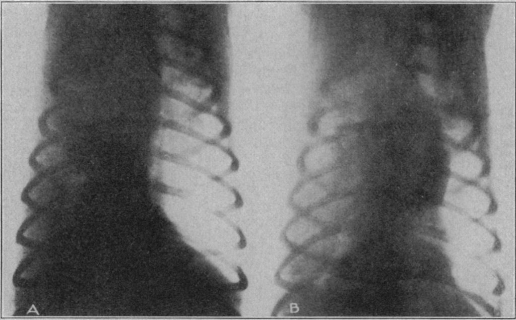
Fig. 5.—A shows unilateral pneumonia in dog PF, twenty-four hours after insufflation of pneumococci type II into the right bronchus; B, restoration of the heart to the normal position and partial clearing of the pneumonic right lung after inhalation of 6 per cent carbon dioxide for two hours. The animal was extremely ill and died two hours later. These pictures are shown in outline in 3 A and of figure 3.
Physiologic evidence in atelectasis
Atelectasis presents a number of puzzling subsidiary problems. Is the causation of atelectasis wholly mechanical, as Coryllos and Birnbaum concluded from their investigations? Or, on the other hand, are there nervous and vascular factors in the collapse of the lung? How are the gases, particularly nitrogen, absorbed? Is there any connection between this problem and that of the spontaneous reinflation of the lung after a therapeutic pneumothorax? Is the difference between pneumothorax and atelectasis wholly dependent on the patency or obstruction of the airways? What light does the evidence from the anatomy of the lung and from the experimental physiology of the bronchial innervation throw on these questions?
The physiologic literature affords little help in understanding these problems. We refer to this literature here chiefly in order to be of service to any reader who may wish to consult it in the hope of finding some hint that others have missed. On the anatomic side the excellent recent review by Macklin (Ref 24) gives a picture of the lung as a contractile organ permeated with nonstriated muscle tissue. This conception is certainly suggestive. On the side of the physiology of the lung the extensive investigations of Carlson and Luckhardt (Ref 25) and the observations of Patterson (Ref 26) show that, if the lungs of men and of frogs were entirely alike, the explanation of atelectasis would probably be found in a nervous effect induced through the vagi and resulting in a prolonged contraction of the intrinsic musculature of the lung. This phenomenon, as described by these investigators in the frog, has not, however, been produced experimentally in a mammal.
The literature on the nervous control of the bronchi in mammals has usually had as its background the phenonmena of asthma, as in the paper of Mount, (Ref 27) and the pharmacologie relief of the symptoms of that disorder; it helps but little toward an understanding of atelectasis. The long papers of Einthoven (Ref 28) and of Beer (Ref 29) are the classics in this field.
The work of Haldane (Ref 30) and his collaborators comes closer to suggestions of importance than that of most other investigators. It shows that shallow breathing in a normal man may leave considerable areas of the lung unventilated, as evidenced by cyanosis. Doubtless a continuation of the experiment and some accumulation of mucus to form a plug in an air tube would produce atelectasis.
Dunn, (Ref 31) and Binger and his collaborators (Ref 32) have demonstrated that multiple small emboli experimentally introduced into the pulmonary circulation may induce shallow breathing. Porter and Newburgh (Ref 33) showed that irritation of the afferent endings of the vagi in the lungs is the cause of the shallow, rapid breathing in pneumonia. Churchill and Cope (Ref 34) gave a similar demonstration for pulmonary congestion and edema. Shallow breathing, according to Meakins, (Ref 35) is in turn the cause of anoxemia in pneumonia, for it ventilates the lungs incompletely. All the factors of a vicious circle are thus provided.
Among the papers dealing with the pharmacologie aspects of the bronchi and their innervation, those of Dixon, (Ref 36) Trendelenburg (Ref 37) and Tiefensee (Ref 38) are especially noteworthy. Reviews giving the physiologic and pharmacologic literature have been published by Starling in Schafer's Textbook of Physiology, (Ref 39) by Boruttau in Nagel's handbook, (Ref 40) Schenck in Tigerstedt's handbook, (Ref 41) and Skramlik (Ref 42) in volume 2 of the excellent handbook now under publication, edited by Bethe and others. Volume 2 of this handbook also contains monographs dealing with the anatomy of the lungs by Felix,(Ref 43) the mechanics of respiration by Rohrer, (Ref 44) the chemistry of the respiratory exchange by Liljestrand,(Ref 45) the pathologic physiology of the airways by Amersbach, (Ref 46) the pathologic physiology of respiration by Hofbaiter (Ref 47) and the pharmacology of respiration by Bayer. (Ref 42)
A survey of all this literature affords, however, little direct evidence that atelectasis can arise directly from nervous influences on the lung. At present, the only clearly demonstrated causes are shallow breathing and the accumulation of mucus, resulting in the mechanical blocking of the air tubes.
Summary and Conclusions
- 1. As the background for this study, the following facts are cited:
(a) If the lungs are not fully distended soon after birth, pneumonia is likely to develop.
(b) After surgical operations, massive, lobar or lobular atelectasis of the lung is a rather frequent occurrence, and is the condition from which postoperative pneumonia develops. This atelectasis or, better, apneumatosis is prevented and relieved, and the risk of pneumonia is eliminated, by the inhalation of carbon dioxide.
(c) The inhalation of 5 per cent carbon dioxide in oxygen, which is now the standard treatment for carbon monoxide asphyxia, is also an effective preventive of postasphyxial pneumonia.
(d) In pneumonia it is the blocking of the lung airways, bronchi or bronchioli, by plugs of thick and sticky secretion which is the critical morbidic factor producing atelectasis and the conditions characteristic of an undrained infection.
- 2. The experiments here reported demonstrate these facts:
(a) Atelectasis that is induced experimentally in dogs by blocking a bronchus is quickly cleared up and the lung is redistended by the deep breathing induced by inhalation of carbon dioxide in proper dilution.
(b) Pneumonia that is induced in dogs by insufflation of a virulent culture of pneumococci is generally overcome, the lung is redistended and the animal is restored to health by inhalation of carbon dioxide sufficient to cause deep breathing and continued until the pneumonic area is cleared.
- 3. Success with this therapy for the relief of pneumonia in patients depends on administering the inhalation as early as possible. If medical pneumonia is thus treated early enough, it appears probable that the results may be as effective as those already attained in postoperative and postasphyxial pneumonia.
Note.—The second paper of this series will deal with the chemical and bactericidal effects of inhalation of carbon dioxide.
Scientific References
| Title | Lobar Pneumonia Considered as Pneumococcic Lobar Atelectasis of the Lung – Full text |
| Journal | Bronchoscopic Investigation, Arch. Surg. 18:190 (Jan.) 1929; Bull. New York Acad. Med. 4:384 (March) 1928. |
| Author | Coryllos, Pol N., and Birnbaum, G. L. |
| Abstract | In a preliminary paper we have given the results of our experimental investigations on lobar pneumonia in dogs, briefly stating the experimental facts on which we have based the theory that lobar pneumonia must be considered as the pneumococcic variety in the large category of obstructive atelectasis of the lung. In the present paper, we propose to develop this theory further and to show that a new light may be thrown on our knowledge concerning lobar pneumonia which may help to explain in a simple and clear way a great number of its features that have heretofore been inexplicable; furthermore, we shall show that pneumonia, up to now considered an exclusively medical disease, may be as much surgical as medical, and as such require an emergency treatment, necessitating a closer collaboration of the thoracic surgeon, the internist and the bronchoscopist. FH. |
| Title | Hyperventilation of the Lungs as a Prophylactic Measure for Pneumonia; The Relief of Experimental Pneumonia; The Production and Relief of Atelectasis of Pneumonia; Symposium on Atelectasis and Pneumonia – Reference |
| Journal | J. A. M. A. 92:434 (Feb. 9) 1929. Proc. Nat. Acad. Sc., Science 69:502 (May) 1929. Am. J. Physiol. 90:317 (Oct.) 1929. |
| Author | Henderson, Y., and Haggard, H. W. Henderson, Y.; Coryllos, P. N.; Haggard, H. W.; Birnbaum, G. L, and Radloff, E. M. Henderson, Y.; Coryllos, P. N.; Holmes, G. W., and Scott, W. J. M. J. A. M. A. 93:96 (July 13) 1929. |
| Abstract | Bigger animals live longer. The scaling exponent for the relationship between lifespan and body mass is between 0.15 and 0.3. Bigger animals also expend more energy, and the scaling exponent for the relationship of resting metabolic rate (RMR) to body mass lies somewhere between 0.66 and 0.8. Mass-specific RMR therefore scales with a corresponding exponent between -0.2 and -0.33. Because the exponents for mass-specific RMR are close to the exponents for lifespan, but have opposite signs, their product (the mass-specific expenditure of energy per lifespan) is independent of body mass (exponent between -0.08 and 0.08). This means that across species a gram of tissue on average expends about the same amount of energy before it dies regardless of whether that tissue is located in a shrew, a cow, an elephant or a whale. This fact led to the notion that ageing and lifespan are processes regulated by energy metabolism rates and that elevating metabolism will be associated with premature mortality–the rate of living theory. The free-radical theory of ageing provides a potential mechanism that links metabolism to ageing phenomena, since oxygen free radicals are formed as a by-product of oxidative phosphorylation.
Despite this potential synergy in these theoretical approaches, the free-radical theory has grown in stature while the rate of living theory has fallen into disrepute. This is primarily because comparisons made across classes (for example, between birds and mammals) do not conform to the expectations, and even within classes there is substantial interspecific variability in the mass-specific expenditure of energy per lifespan. Using interspecific data to test the rate of living hypothesis is, however, confused by several major problems. For example, appeals that the resultant lifetime expenditure of energy per gram of tissue is ‘too variable’ depend on the biological significance rather than the statistical significance of the variation observed. Moreover, maximum lifespan is not a good marker of ageing and RMR is not a good measure of total energy metabolism. Analysis of residual lifespan against residual RMR reveals no significant relationship. However, this is still based on RMR. A novel comparison using daily energy expenditure (DEE), rather than BMR, suggests that lifetime expenditure of energy per gram of tissue is NOT independent of body mass, and that tissue in smaller animals expends more energy before expiring than tissue in larger animals. Some of the residual variation in this relationship in mammals is explained by ambient temperature. In addition there is a significant negative relationship between residual lifespan and residual daily energy expenditure in mammals. A potentially much better model to explore the links of body size, metabolism and ageing is to examine the intraspecific links. These studies have generated some data that support the original rate of living theory and other data that conflict. In particular several studies have shown that manipulating animals to expend more or less energy generate the expected effects on lifespan (particularly when the subjects are ectotherms). However, smaller individuals with higher rates of metabolism live longer than their slower, larger conspecifics. An addition to these confused observations has been the recent suggestion that under some circumstances we might expect mitochondria to produce fewer free radicals when metabolism is higher–particularly when they are uncoupled. These new ideas concerning the manner in which mitochondria generate free radicals as a function of metabolism shed some light on the complexity of observations linking body size, metabolism and lifespan. Prior to the introduction of the treatment now prevalent for carbon monoxide asphyxia by means of inhalation of carbon dioxide and oxygen,1 pneumonia was one of the recognized sequels of severe but not immediately fatal gassing.2 It is therefore a suggestive fact that, among cases treated by inhalation of carbon dioxide, postasphyxial pneumonia does not occur. Among hundreds of cases thus treated, of which we have records, there is not a single report of a subsequent pneumonia.In this laboratory a few years ago it was found also, as previously observed by Oliver,3 that dogs after prolonged asphyxia may develop a condition of acute congestion of the lungs. Against this condition in dogs, inhalation of carbon dioxide in oxygen is an effective prophylaxis. These facts, when they first came to our attention, were both unforeseen and unexplainable. Little stress was laid on them in the publicationsBigger animals live longer. The scaling exponent for the relationship between lifespan and body mass is between 0.15 and 0.3. Bigger animals also expend more energy, and the scaling exponent for the relationship of resting metabolic rate (RMR) to body mass lies somewhere between 0.66 and 0.8. Mass-specific RMR therefore scales with a corresponding exponent between -0.2 and -0.33. Because the exponents for mass-specific RMR are close to the exponents for lifespan, but have opposite signs, their product (the mass-specific expenditure of energy per lifespan) is independent of body mass (exponent between -0.08 and 0.08). This means that across species a gram of tissue on average expends about the same amount of energy before it dies regardless of whether that tissue is located in a shrew, a cow, an elephant or a whale. This fact led to the notion that ageing and lifespan are processes regulated by energy metabolism rates and that elevating metabolism will be associated with premature mortality–the rate of living theory. The free-radical theory of ageing provides a potential mechanism that links metabolism to ageing phenomena, since oxygen free radicals are formed as a by-product of oxidative phosphorylation. Despite this potential synergy in these theoretical approaches, the free-radical theory has grown in stature while the rate of living theory has fallen into disrepute. This is primarily because comparisons made across classes (for example, between birds and mammals) do not conform to the expectations, and even within classes there is substantial interspecific variability in the mass-specific expenditure of energy per lifespan. Using interspecific data to test the rate of living hypothesis is, however, confused by several major problems. For example, appeals that the resultant lifetime expenditure of energy per gram of tissue is ‘too variable’ depend on the biological significance rather than the statistical significance of the variation observed. Moreover, maximum lifespan is not a good marker of ageing and RMR is not a good measure of total energy metabolism. Analysis of residual lifespan against residual RMR reveals no significant relationship. However, this is still based on RMR. A novel comparison using daily energy expenditure (DEE), rather than BMR, suggests that lifetime expenditure of energy per gram of tissue is NOT independent of body mass, and that tissue in smaller animals expends more energy before expiring than tissue in larger animals. Some of the residual variation in this relationship in mammals is explained by ambient temperature. In addition there is a significant negative relationship between residual lifespan and residual daily energy expenditure in mammals. A potentially much better model to explore the links of body size, metabolism and ageing is to examine the intraspecific links. These studies have generated some data that support the original rate of living theory and other data that conflict. In particular several studies have shown that manipulating animals to expend more or less energy generate the expected effects on lifespan (particularly when the subjects are ectotherms). However, smaller individuals with higher rates of metabolism live longer than their slower, larger conspecifics. An addition to these confused observations has been the recent suggestion that under some circumstances we might expect mitochondria to produce fewer free radicals when metabolism is higher–particularly when they are uncoupled. These new ideas concerning the manner in which mitochondria generate free radicals as a function of metabolism shed some light on the complexity of observations linking body size, metabolism and lifespan. |
| Title | Diseases of Infancy and Childhood, part 2, ed. 8, Chapters 1 and 2 – Full text |
| Journal | New York, D. Appleton & Company, 1922. |
| Author | Holt, L. E., and Howland, J. |
| Abstract | The first edition of this volume by the late L. Emmett Holt appeared in 1897. Subsequent editions were revised by John Howland; the present edition was prepared by L. Emmett Holt Jr. and Rustin McIntosh. In the present revision the authors have endeavored as much as possible to retain the personal opinions and attitudes of the original authors. They supplement the text with a bibliography and add much material on pathogenesis. So extensive have been advances in the field of nutrition, the blood and allergy and also on some of the infectious diseases and on endocrinology that certain sections have been rewritten completely and extensive revisions made in others. Perhaps a dozen associates have been called on by the authors for the revision of some of the special sections of this book. Its usefulness is manifest from its record of sales. |
| Title | The Prevention and Treatment of Asphyxia in the New- Born – Reference |
| Journal | J. A. M. A. 90:583 (Feb. 25) 1928. |
| Author | Henderson, Y. |
| Abstract | The first quarter of an hour after birth is the most dangerous period of life. Its mortality is as great as that of any subsequent month. No single discovery in medical science or improvement in practice could do more to save lives than would measures to avoid the losses that now occur within a few minutes after birth. There are probably no procedures in medical practice which are so nearly the same as those of the dark ages, or which have as yet been influenced so little by modern scientific knowledge, as those of the midwife or obstetrician in dealing with a nonbreathing new-born child. Perhaps an excuse may be found in the statements still contained in textbooks of physiology and obstetrics regarding the questions why the fetus does not breathe in the uterus, and why it does start to breathe after birth. This topic is generally discussed rather philosophically. |
| Titel | Postoperative Pneumonia – Full text |
| Journal | Arch. Surg. 18:2167 (May) 1929. |
| Author | Smith, M. K., and Morton, P. C. |
| Abstract | Postoperative pneumonia, despite the study it has received, remains a dreaded complication of surgical therapy. In an assembled table of figures from clinics here and abroad, Cutler and Hunt1 found that in operations on 41,368 patients there was an incidence of pneumonia of 1.12 per cent, 46 per cent of whom died—a mortality by operation of about 0.5 per cent. These figures require explanation, however. The apparently low general mortality of 0.5 per cent is greatly increased in certain operations, notably those on the stomach, as will be discussed later. On the other hand, the high death rate in those cases in which pneumonia develops is due to a considerable extent to the association of other grave conditions. Among patients who were well and came in for an operation of choice and then developed pneumonia Whipple2 found only nine deaths in sixty-one cases. |
| Title | Postoperative Massive Atelectasis; Postoperative Pneumonia; Prophylaxis of Postoperative Pneumonia: Preliminary Report of Some Experiments After Upper Abdominal Operations – Full text |
| Journal | J. A. M. A. 90:1759 (June 2) 1928. J. A. M. A. 8:384 (Feb. 2) 1924. Anesth. & Analg. 7:187 (May-June) 1928. |
| Author | Scott, W. J. M., and Cutler, E. C. Elwyn, H. Sise, L. F.; Mason, R. L., and Bogan, I. K. |
| Abstract | In a previous paper, 1 evidence was presented which indicated that the posture of the patient both during the operation and in the early postoperative period influenced the localization of postoperative massive atelectasis. It is our purpose in this paper to study the influence of variations in respiratory volume on the development of postoperative massive atelectasis. There are now available excellent papers covering the incidence and the clinical syndrome of this condition. Many etiologic factors have been proposed but no primary cause has been found. We ourselves believe that the process is initiated by a nervous reflex, probably largely vasomotor, which results in a narrowing of the lumen in the peripheral bronchioles by venous engorgement, swelling of the mucous membrane, and the elaboration of a tenacious secretion. Whether a common factor always initiates the reflex is uncertain, and we have felt that several secondary factors contributed to the completion |
| Title | Apparatus zur Kohlensaureinhlation und bildliche Darstellung der Kohlensaureanwendung am Kranken |
| Journal | Deutsche Ztschr. f. Chir. 205:22, 1927 |
| Author | Dzialoszynski, A. |
| Title | Prevention of Postoperative Lung Complications by Inhalation of Carbon Dioxide |
| Journal | Zentralbl. f. Gynak. 52:2010 (Aug. 11) 1928. |
| Author | Fisher, E. |
| Title | Principles and Practice of Medicine, revised by Thomas McCrae – Full text |
| Journal | Ed. 10, New York, D. Appleton & Company, 1926, p. 659. |
| Author | Osler, W. |
| Title | Ueber eine Methode zur experimentellen Erzeugung von Pneumonie und ueber einige mit dieser Methode erzielte Ergebnisse, Berl. klin. Wchnschr |
| Journal | July 20, 1914, p. 1351. |
| Author | Meltzer, S. J |
| Title | Fatal Apnoea and the Shock Problem |
| Source | Bull. Johns Hopkins Hosp. 21:235 (Aug.) 1910. |
| Author | Henderson, Y. |
| Title | Administration of Carbon Dioxide After Anesthesia and Operation; Acapnia Theory |
| Journal | J. A. M. A. 76:672 (March 5) 1921. J. A. M. A. 77:424 (Aug. 6) 1921. |
| Author | Henderson, Y. Haggard, H. W., and Coburn, R. C. |
| Title | The Treatment of Carbon Monoxide Asphyxia by Means of Oxygen Plus Carbon Dioxide Inhalation; Resuscitation from Carbon Monoxide Asphyxia from Ether or Alcohol Intoxication, and from Respiratory Failure Due to Other Causes; Noxious Gases – Reference |
| Journal | J. A. M. A. 79:1137 (Sept. 30) 1922. J. A. M. A. 83:758 (Sept. 6) 1924. Chemical Catalog Co., 1925. |
| Author | Henderson, Y., and Haggard, H. W. |
| Abstract | At a meeting of the Commission on Resuscitation from Carbon Monoxid Asphyxia on Nov. 15, 1921, the writers were appointed a subcommittee to conduct investigations both in the field and in the laboratory on the treatment of carbon monoxid asphyxia by the inhalational method, to be here described. There is already available a considerable mass of theoretical knowledge and experimental material on which to base further advance along the line indicated. Recent investigations in this laboratory1 have confirmed the correctness of the view that carbon monoxid has no direct toxic action on the brain, or other organs or tissues of the body, except only the blood. It acts wholly through its combination with the hemoglobin or red coloring matter of the blood. When so combined, the hemoglobin is for the time deprived of the power to transport oxygen from the lungs to the tissues of the body. |
| Title | Carbon Monoxide Asphyxia: The Problem of Resuscitation |
| Journal | J. Indust. Hyg. 4:463, 1922-1923. |
| Author | Drinker, C. K., and Cannon, W. B. |
| Title | Quoted from a report on autopsies |
| Journal | 1922, Department of Pathology, Yale Medical School. |
| Author | Dr. R. A. Lambert |
| Title | Massive Collapse of the Lung – Reference |
| Journal | Lancet 2:1351, 1908 and 2:1080, 1910. |
| Author | Pasteur, W. |
| Title | Diseases of the Bronchi, Lungs and Pleura |
| Journal | Philadelphia, Lea & Febiger, 1915. |
| Author | Mendelsohn, A., quoted by Lord, F. F. |
| Title | Arch. f. exper. Path. u |
| Journal | Pharmakol. 10:54, 1879. |
| Author | Lichtheim, L. |
| Title | Obstructive Massive Atelectasis of the Lung – Reference |
| Journal | Arch. Surg. 16:501 (Feb.) 1928. |
| Author | Coryllos, Pol N., and Birnbaum, G. L. |
| Abstract | Pasteur described postoperative atelectasis or collapse of the lung as a condition of this organ characterized by "the total deflation (as opposed to lobular or patchy collapse) of a large area of lung tissue, of sudden onset—in the absence of any signs of obstruction of the air ways or of any known source of compression—due to failure of inspiratory power and attended by definite physical symptoms and signs." Since then, and even long before Pasteur had again brought it up as an actuality, many papers have been written on this puzzling condition. None seems to have given a definite solution to the etiology and the mechanism of it. In this paper, we propose to give the results of our experimental work conducted on fifty-six dogs. A careful study of the clinical symptoms, the physiologic phenomena, the roentgen-ray observations on serial pictures and the pathologic and bacteriologic condition of our animalsattern. |
| Title | Displacement of Heart in Pneumonia in Childhood – Full text |
| Journal | Arch. Dis. Childhood 4:155 (Aug.) 1929, quoted in J. A. M. A. 93:1099 (Oct. 5) 1929. |
| Author | Tallerman, K. H., and Jupe, M. H. |
| Title | Experimental Studies on Denervated Lungs – Reference |
| Journal | Arch. Surg. 16:1153 (June) 1928. |
| Author | Fontaine, R., and Herrmann, L. G. |
| Abstract | There is no vital structure in the body that is so frequently the seat of disease as the lung; still the practical importance of the innervation of this organ is not generally appreciated. The complexity and often the contradictory evidence regarding this innervation have prevented the real facts from being disseminated even in medical schools. Recent evidence, however, emphasizes the important rôle which the nerves that lead to the lung may play in clinical disorders. Binger and his associates1 have shown that the tachypnea which accompanies acute pulmonary disease in the absence of anoxemia is of nervous origin. The cause of sudden death in cases of pulmonary embolism and following thoracentesis, as well as the cause of bronchial asthma and allied conditions, may also be nervous in origin. It has also been suggested that postoperative massive atelectasis (collapse) of the lungs may, in part, be due to a disturbed |
| Title | The Musculature of the Bronchi and Lungs – Reference |
| Journal | Physiol. Rev. 9:1 (Jan.) 1929. |
| Author | Macklin, C. C. |
| Title | Studies on the Visceral Sensory Nervous System – Reference |
| Journal | Am. J. Physiol. 54:55 (Nov.) 1920; 55:13 and 31 (Feb.), 212 (March), 366 (April); 56:72 (May); 57:299 (Sept.) 1921. |
| Author | Carlson, A. J., and Luckhardt, A. B. |
| Title | Studies on the Visceral Sensory Nervous System: IX. The Readjustment of the Peripheral Lung Motor Mechanism After Bilateral Vagotony in the Frog – Reference |
| Journal | Am. J. Physiol. 58:169 (Nov.) 1921. |
| Source | Patterson, T. L. |
| Title | Experimental Study of the Effects of Stimulation and Section of the Vagal Innervation to the Bronchi, and Their Possible Relation to Asthma |
| Journal | Am. J. M. Sc. 177:697 (May) 1929. |
| Author | Mount, H. T. R. |
| Title | Ueber die Wirkung der Bronchialmuskeln, nach einer neuen Methode untersucht und uber Asthma nervosum |
| Journal | Arch. f. d. ges. Physiol. 51:367, 1892. |
| Author | Einthoven, W. |
| Title | Ueber den Einfluss der peripheren Vagusreizung auf die Lunge |
| Journal | Arch. f. Anat. u. Physiol., 1892, p. 101. |
| Author | Beer, Theodor |
| Title | Respiration – Full Text |
| Journal | New Haven, Conn., Yale University Press, 1922, p. 136. |
| Author | Haldane, J. S. |
| Title | Experimental Studies on Rapid Breathing: II. Tachypnea, Dependent upon Anoxemia, Resulting from Multiple Emboli in the Larger Branches of the Pulmonary Artery – Reference |
| Journal | J. Clin. Investigation 1:155 (Dec.) 1924. Binger, A. L., and Moore, R. L.: J. Exper. Med. 45:655, 1927. |
| Author | Binger, A. L. |
| Title | Am. J. Physiol. |
| Journal | 42:175, 1916; 43: 455, 1917. J. Exper. Med. 24:583, 1916. |
| Author | Porter, W. T., and Newburgh, L. H. Porter; Newburgh and Means, J. H. |
| Title | The Rapid Shallow Breathing Resulting from Pulmonary Congestion and Edema – Reference |
| Journal | J. Exper. Med. 49:531, 1929. |
| Author | Churchill, E. D., and Cope, O. |
| Abstract | These experiments record the effects of the experimental production of pulmonary congestion and edema in a lung completely isolated from the general circulation, but with an intact nerve supply. The resulting changes are: a slowing of the heart rate, a fall in systemic blood pressure and a temporary inhibition of respiration succeeded by rapid shallow breathing. The pulse rate and blood pressure show a rapid and spontaneous return to initial conditions. The respirations show a partial but not a complete return to their former rate and depth. The effects on respiration are similar to those described by Dunn and Binger and Moore which follow multiple embolism of the pulmonary circuit with starch granules. The alterations in the pulse rate and blood pressure are characteristic of the effects of vagal stimulation. A chemical effect on the respiratory center is excluded by the nature of the preparation. These results, therefore, add further evidence to support the hypothesis that the rapid shallow breathing attending congestion and edema of the lungs is due to the stimulation of nerve endings in the lungs. |
| Title | Harmful Effects of Shallow Breathing with Special Reference to Pneumonia – Reference |
| Journal | Arch. Int. Med. 25:1 (Jan.) 1920. |
| Author | Meakins, J. |
| Abstract | It has been demonstated by Haldane, Meakins and Priestley1 that abnormal shallowness of the respirations produces anoxemia. This is demonstrated by the occurrence of periodic breathing, cyanosis and other symptoms indicative of this condition. The manner in which this abnormal type of breathing produces the anoxemia has also received considerable attention from them. Keith has demonstrated that the lungs do not expand after the manner of the bladder when external pressure is reduced, but like a Japanese fan. He has also pointed out that expansion of the lungs does not take place constantly and uniformly throughout. Therefore this type of breathing would of necessity exaggerate the uneven distribution of air to the alveoli. |
| Title | Contributions to the Physiology of the Lungs: I. The Bronchial Muscles, Their Innervations and the Action of Drugs upon Them, Bronchodilator Nerves, Studies in the Pulmonary Circulation: I. The Vaso-Motor Supply, Studies in the Pulmonary Circulation: II. The Action of Adrenaline and Nicotine – Reference |
| Journal | J. Physiol. 29:97, 1903. J. Physiol. 45:413, 1913. J. Physiol. 65:299 (June 24) 1928. J. Physiol. 67:77 (Feb. 28) 1929. |
| Author | Dixon, W. E., and Ransom, F. Dixon, W. E., and Hoyle, J. C. Dixon, W. E., and Hoyle, J. C. |
| Title | Physiologische und pharmakologische Untersuchungen an der isolierten Bronchialmuskulatur, Versuche an der isolierten Bronchialmuskulatur |
| Journal | Arch. f. exper. Path. u. Pharmakol. 69:79, 1912. Zentralbl. f. Physiol. 26:1, 1913. |
| Author | Trendelenburg, P. |
| Title | Pharmakologische Studien auf der Bronchialmuskulatur: I. Methodik, II. Ueber die Bedeutung der Blutbeschaffenheit fur den Tonus der Bronchialmuskeln und ihr |
| Journal | Ansprechen auf Gifte 139:139, 1929. |
| Author | Tiefensee, K. |
| Title | The Muscular and Nervous Mechanism of the Respiratory Movements |
| Journal | Text-book of Physiology, vol. 2, New York, The Macmillan Company, 1900, p. 274. |
| Author | Starling, E. H. |
| Title | Die Atembewegungen und ihre Innervation: IV |
| Journal | Die Innervation der Atembewegungen, in W. Nagel: Handbuch der Physiologie des Menschen, Braunschweig, F. Vieweg und Sohn, 1909, vol. 1, p. 29. |
| Author | Boruttau, Heinrich |
| Title | Atembewegungen |
| Journal | Handbuch der physiologischen Methodik, vol. 2, Leipzig, 1908. |
| Author | Schenck, F. |
| Title | Die Physiologie der Luftwege |
| Journal | Handbuch der normalen und pathologischen Physiologie, ed. by Bethe, Bergmann Embden and Ellinger, Berlin, Julius Springer, 1925, vol. 2, p. 128. |
| Author | Von Skramlik, Emil |
| Title | Anatomie der Atmungsorgane |
| Journal | Handbuch der normalen und pathologischen Physiologie, vol. 2, p. 37. |
| Author | Felix, Walther |
| Title | Physiologie der Atembewegung |
| Journal | Handbuch der normalen und pathologischen Physiologie, vol. 2, p. 70. |
| Author | Rohrer, Fritz |
| Title | Chemismus des Lungengaswechsels |
| Journal | Handbuch der normalen und pathologischen Physiologie, vol. 2, p. 190. |
| Author | Liljestrand, G. |
| Title | Patho-Physiologie der Luftwege |
| Journal | Handbuch der normalen und pathologischen, Physiologie, vol. 2, p. 307. |
| Author | Amersbach, K. |
| Title | Pathologische Physiologie der Atmung |
| Journal | Handbuch der normalen und pathologischen Physiologie, vol. 2, p. 337. |
| Author | Hofbauer, Ludwig |
| Title | Pharmakologie der Atmung |
| Journal | Handbuch der normalen und pathologischen Physiologie, vol. 2, p. 455. |
| Author | Bayer, Gustav |


